


Advanced imaging and cytometry laboratory
Key personnel:
dr Michał Majkowski
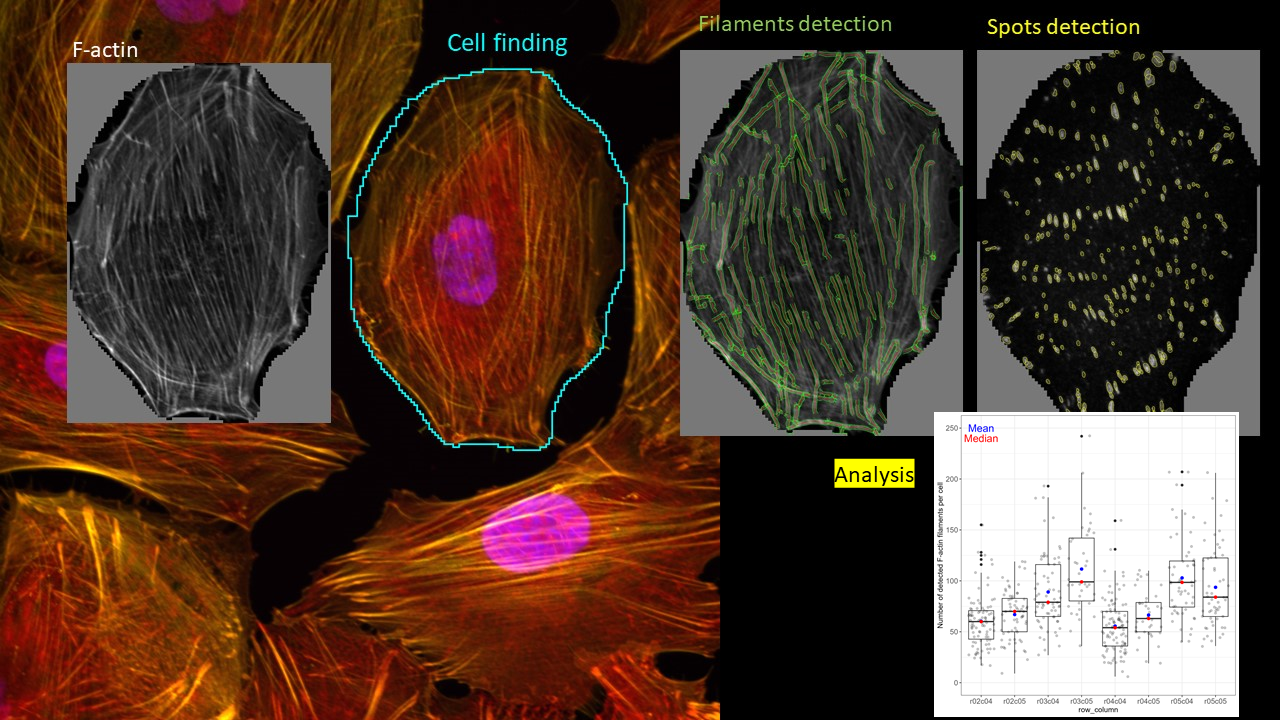
Image courtesy of Magdalena Kot, Aleksandra Simiczyjew, Michał Majkowski.
Equipment:
Confocal microscope Stellaris 8 with FLIM and STED modules
Research confocal microscope. FLIM is an imaging technique in which each pixel contain information not only about fluorescence signal intensity but also about fluorescence lifetime. Measuring the latter might provide information about molecular environment of the probe. This technique is also useful in FRET measurement. STED uses 660 nm depletion laser line and is combined with fluorescence lifetime measurement. This allow – in theory – to achieve resolution in XY in the region of 70-80 nm. The system also offers FCS measurements.
Dry, water- and oil-immersion optics, incubation chamber with CO2. Wide range of excitation lines and great flexibility in detection range.
Elyre 7 microscope
Research super-resolution microscope. Uses standard samples stained with the most popular fluorophores and employs structured pattern of illumination to achieve resolution in the region of 70 nm. Also offers TIRF, STORM and PALM imaging. Dry, water- and oil-immersion optics, incubation chamber, 405 nm, 488 nm, 561nm, 633 nm laser lines. Two cameras: sCMOS and back-thinned EMCCD. Resolution in Z in the region of 300 nm. Fluorescence filters for four typical fluorofores: DAPI (or similar), Cy2 (or similar), Cy3 (or similar) and Cy5 (or similar).
Opera Phenix HTS/HCS microscope
Spinning disc based confocal microscope, optimized for large scale imaging, is able to produce vast number of images in relatively short time. Equipped with robotic arm that shuttles samples between incubator/plate rack and microscope. An injector allows to add a compound of interest to the sample while capturing images what allows to image very fast processes. Harmony software does smooth image processing and quantification. The system also allows high scale FRET imaging between CFP and YFP.
The core also offers low and high scale image quantification in FIJI software.
Lab is also equipped with FACS and flow cytometer.
Contact: michal.majkowski@uwr.edu.pl
The purchase of the equipment was financially supported by the Research Equipment Fund (FAB) of the „Excellence Initiative — Research University” (IDUB) program for the University of Wroclaw.